Artificial Intelligence In Ophthalmology Center
The UIC Artificial Intelligence in Ophthalmology (Ai-O) is a center of excellence for Artificial Intelligence research, theory, applications, and education in ophthalmology.
The Artificial Intelligence in Ophthalmology Center Heading link
Overview Heading link
Vision
As we amass more data, the greatest advances in ophthalmic healthcare solutions of the 21st century will be the creation of tools that can utilize and integrate diverse ophthalmic imaging, clinical, genetic, and socio-demographic data for complex AI solutions.
Mission
Our mission is to develop Human Centered-AI for patient care in an interpretable, ethical and reproducible manner.
Cores
- Theory and Learning
- Medical Imaging
- Clinical Research
- Translational
- Public Health and Education
Ai-O Center Heading link
Located at the Eye and Ear Infirmary (EEI)
Select Projects Heading link
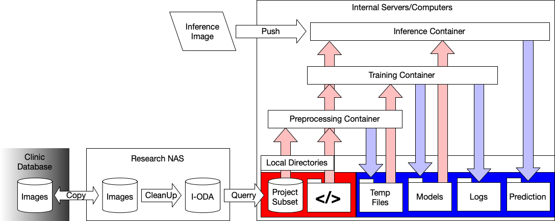
I-ODA
I-ODA is a live databank that includes real-world longitudinal clinical ophthalmic images and patient metadata, with the purpose of advancing state-of-the-art computer vision applications in ophthalmology, and improving upon the translatable capacity of machine learning based applications.
Project lead: Joelle Hallak
Team: Nooshin Mojab, Abdullah Aleem, Manoj Prabhakar Nallabothula, Joe Baker, Darvin Yi
Auto Ptosis
Auto Ptosis is a pipeline for detecting and calculating the features to help diagnosis ptosis. This disease presents itself with the “drooping” or “falling” of the upper eyelid and can lead to an impairment of visual field. We tackle this task through several different levels of hand-crafted design: (1) we train a classification network to differentiate healthy and ptosis eyes in an end-to-end fashion, (2) we use keypoint detection networks to delineate eyelids and pupils to derive the MRD1 distance (the marginal reflex distance-1, the gold standard of ptosis diagnosis), and (3) we also calculate supporting features such as percentage of iris visible through the eyelids. Features such as the latter were too difficult to calculate before, but with machine learning, can be an automatic output from any front-facing photograph. Calculating novel metrics like these can help expand clinical workflow and eliminate the need for complex clinical imaging set-ups, potentially supporting efforts in telemedicine.
Project lead: Pete Setabutr
Team Lead: Darvin Yi
Team: Abdullah Aleem, Manoj Prabhakar Nallabothula
Another selected project Heading link
AI-Utils
The AI Utils project aims to create basic deep learning AI tools for internal research in UIC’s department of ophthalmology and bioengineering. This is done through an amalgamation of API’s, protocols, and software packages hosted in the AI Utils repository . Through dockering workflows, we will define potential folder hierarchies and easy to load models, based on current state of the art (SOTA) performances. For new projects, users will have to simply format their data according to AI Utils protocol, and training should become as simple as a single line command. Upon training a network, AI Utils will also provide an API for future inference calls through docker as well, which will enable easy deployment on cloud compute clusters or even our own backend servers. By well defining and documenting agreements between users and developers, we can strive to democratize AI.
Project lead: Darvin Yi
Team: Abdullah Aleem, Manoj Prabhakar Nallabothula, Joelle Hallak
Our Team Heading link
-
Individuals in leadership roles at the center
Darvin Yi
Co-Executive DirectorThasarat Sutabutr Vajaranant
Director of Data Sourcing and StrategyR.V. Paul Chan
Co-Founder, Co-Executive DirectorDennis Edmonds
Technical Director -
Individuals who advise the center
Joelle Hallak
Advisor for Real World Data Science and Co-FounderEmail:
Pete Setabutr
Advisor for Oculoplastics SciencePhone:
-
Students past and present
Jacob Love
Jacob Love is a master’s student in Computer Science at the University of Illinois Chicago (UIC) and an artificial intelligence researcher at the Artificial Intelligence in Ophthalmology (Ai-O) Center at UIC. Previously he received undergraduate degrees in Cellular-Molecular Neuroscience and Physics from the Loyola University of Chicago, where he discovered a passion for computer science while working in a circadian biology lab.
Jacob’s research interests involve using artificial intelligence to aid in understanding and operating complex systems. His current research includes developing computer vision algorithms for advancing clinical ophthalmology and theoretical work furthering gradient descent methods for multiagent systems. Outside of research, his hobbies include drawing, reading, and listening to music.
Simon (Jiechao) Ma
Simon is a Ph.D. student in the Biomedical Engineering department. His current research focuses on investigating deep neural networks for abnormality detection and disease classification in ophthalmic images.Born in Guangzhou (also known as Canton), China, Simon received his bachelor’s and master’s degree in Mechanical Engineering at China Agricultural University, and a PhD in Agricultural and Biological Engineering from Purdue University. He loves playing table tennis, soccer, and 3d printing.
Rogerio Garcia Nespolo
Rogerio Nespolo is a third-year Ph.D. student in Biomedical Engineering with a research appointment at the UIC Department of Ophthalmology & Visual Sciences. His work comprises surgical guidance via computer vision and surgical data science for ophthalmic procedures.
Homa Rashidisabet, PhD
Homa is a Ph.D. student in the Biomedical Engineering department at the University of Illinois at Chicago (UIC). Her doctoral research focuses on developing predictive models for abnormality detection in real-world ophthalmic images via deep learning (e.g., predicting glaucoma disease from Fundus images), as well as developing predictive models for digital phenotyping from real-world sensor data (e.g., predicting depression relapse from actigraphy data via deep-learning-based multivariate timeseries anomaly detection methods).
She is interested in using AI to transform medicine from a reactive to a proactive discipline.
Homa holds a bachelor’s degree in Applied Mathematics from the University of Tehran in Tehran, Iran. She enjoys swimming, singing, studying astronomy, and ancient history.
Abhishek Sethi
Abhishek is a second year medical student interested in the intersection between computer science, engineering, and medicine. My research interests include computer vision, natural language processing, and robotics.
Mahtab Faraji
Mahtab is currently pursuing her Ph.D. in Biomedical Engineering at the University of Illinois at Chicago (UIC). After completing a Bachelor’s degree in Biomedical Engineering from Sahand University of Technology, Iran, in 2014, she completed her Master’s degree in the same field at Iran University of Science and Technology, in 2018. Her research passion lies in applying Artificial Intelligence (AI) methods to enhance computer-aided diagnosis, with a primary emphasis on Ophthalmology and Cardiovascular medicine. In her free time, she enjoys social activities, running, and dancing.
Alumni
- Minhaj Nur Alam, PhD
- Abdullah Aleem, MS
- Sasha Kravets, PhD
- Nooshin Mojab, PhD
- Kate Romond