Research Programs
The Division of Pathology Research is organized in a matrix of programs and individual laboratories. Our research community is collaborative and multi-disciplinary, encompassing not only the research and clinical faculty within the Pathology Department, but also other departments throughout the UIC College of Medicine, the University of Illinois, and outside institutions.
Research Programs Heading link
-
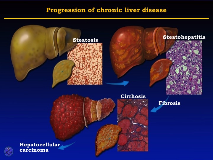
Fostering innovation and collaboration in liver research
Four pressing concerns drive research on liver diseases at UIC: the escalation in chronic liver disease, the rapid growth of alcoholic and non-alcoholic steatohepatitis worldwide, the increasing incidence of liver cancer and the role of the gut-liver interaction in liver disease. Basic, translational and clinical research at UIC is geared toward finding solutions.
The Liver Program in the Department of Pathology at UIC has brought together an exceptional team of multidisciplinary investigators to study the multiple aspects of liver disease and apply new technologies discovered by basic research to the medical care of patients with liver disease. Through this mission our Program encourages and supports important biomedical research into liver disease; addressing major local, national and international needs, through a cohesive group of researchers from different disciplines involved in liver research.
The Liver Program members are themselves quite diverse. They represent more than 10 departments at UIC and nearby institutions, including the Jesse Brown VA Medical Center and Rush University.
The objectives of the Liver Program are to:
- Apply new technologies discovered by basic research to the medical care of patients with liver disease,
- Form a cohesive group of investigators from different disciplines involved in liver research,
- Foster excellence and scientific collaboration among its members,
- Recruit new investigators to the field of hematology, providing a community platform for the exchange of ideas,
- Enhance national and international recognition of the faculty in the Liver Program involved in both basic and clinical research in liver biology and pathology.
- Increase funding for liver research at the University of Illinois at Chicago.
Research themes
The overall focus of the Center is on the integrative biology of the liver. Within this framework, the research performed by individual members is quite diverse, ranging from studies of basic liver cell biology to the investigation of liver physiology and pathophysiology in animal models and humans to the evaluation of treatment practices and outcomes for patients with liver disease. The main themes are:
- Liver injury and repair
- Hepatic physiology and metabolism
- Liver fibrosis
- Alcoholic liver disease
- Non-alcoholic liver disease
- Hepatocellular carcinoma
- Hepatitis C
- Hepatitis B
- Tissue imaging
- Cholesterol fluxes
- Gut microbiome
Related Resources:
-
Innovative Diagnostics
Our goal is to bring the latest innovations in scientific and technical advances to the development of new, patient-oriented diagnostic testing.
Current areas of interest in new diagnostic test development and discovery include:
Brain tumor molecular diagnostics
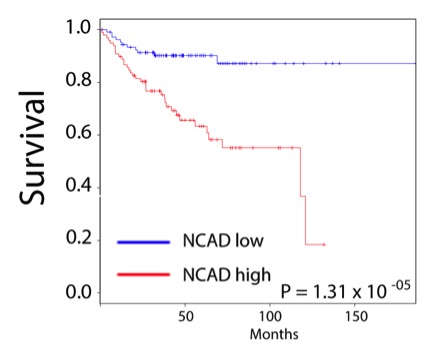
We are developing highly specific and sensitive methods to detect and quantify mutations that can predict the outcome of adult and pediatric brain tumors. Ongoing work has identified a cell adhesion molecule, CDH11 in prognosis of glioblastoma. In medulloblastoma, the most common malignant pediatric tumor, we have identified a gene signature that predicts that a subset of Sonic Hedgehog tumors is characterized by overactivity of the N-cadherin cell adhesion molecule.
Faculty:
- Tibor Valyi-Nagy, MD, PhD
- Peter Gann, MD, PhD
- Shrihari Kadkol, MD, PhD
Prostate Cancer
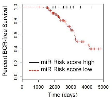
Overly aggressive treatment of indolent prostate cancer (PCa) occurs because identification of patients with a low-risk of having aggressive disease remains challenging. We have developed a “signature” of microRNA’s that is present in patient serum of men with prostate cancer that predicts whether their disease shows aggressive features. Ongoing work utilized computational and molecular approaches to refine the signature and validate its effectiveness on large patient cohorts.
Faculty:
- Larisa Nonn, PhD
- Peter Gann, MD, PhD
Minimally-Invasive Breast Carcinoma Monitoring
Tumor-derived DNA can be detected in the systemic circulation in the form of cell-free plasma DNA (cfpDNA), and the ability to interrogate tumor-derived DNA from an easily acquired biospecimen like cfpDNA has practical advantages over analysis of tissue specimens. Furthermore, cfpDNA has practical advantages over analysis of tissue species. Furthermore, cfpDNA can be monitored serially and could provide a dynamic measure of tumor profession and response to therapy. Activating mutations in the PIK3CA gene represent the most common molecular aberration in luminal breast cancers. We have developed a highly sensitive digital PCR method to detect and quantify levels in patient serum of the most common “hotspot” mutations in the PIK3CA gene in patients with breast cancer (abstract). Ongoing work will further refine the methods and validate the clinical utility of this approach in monitoring breast cancer progression and response to treatment.
Faculty:
- Kent Hoskins, PhD
-
Neurovirology and Ocular Virology
Research program activities focus on mechanisms of herpes simplex virus infection (HSV)-induced nervous system and ocular disease, potential role of HSV infection in the pathogenesis of neurodegenerative diseases including Alzheimer disease, molecular mechanisms of HSV entry into cells, mechanisms of tumor resistance against HSV-mediated oncolytic virotherapy and development of new anti-viral agents.
Faculty
- Tibor Valyi-Nagy, MD, PhD
-
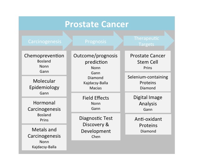
Prostate
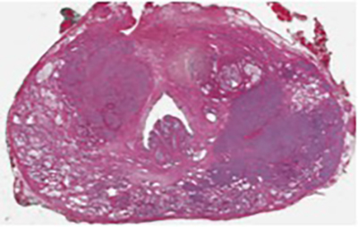
Prostate research is a focus area for several Pathology faculty members. This research addresses knowledge gaps in prostate cancer prevention, prognostic biomarkers of aggressive cancers and understanding of potential therapeutic targets. Our translational approach to research maintains clinical relevance and focus on the prostate cancer patient, while also utilizing in vitro and in vivo models to reveal molecular mechanisms. Funded projects range from clinical trials to molecular next-generation sequencing.
Faculty
Faculty:
- Maarten C. Bosland, DVSc, PhD
- Prostate cancer chemoprevention
- Hormonal carcinogenesis of the prostate & endocrine disruptors
- Alan M. Diamond, PhD
- The role of anti-oxidant proteins in prostate cancer etiology
- The molecular and biological consequences of allelic variations in the genes for selenium-containing proteins
- Peter H. Gann, MD, ScD
- Prostate cancer biomarker development
- Molecular epidemiology of prostate cancer
- Digital microscopy and image analysis for prostate cancer prognosis
- Andre Kajdacsy-Balla, MD, PhD
- Prostate cancer clinical outcomes prediction methods
- The effect of environmental agents on prostate cancer progression and metastasis
- Virgilia Macias, MD
- Prognostic markers in prostate cancer
- Larisa Nonn, PhD
- Zinc homeostasis alterations in prostate carcinogenesis
- microRNAs as prognostic markers of cancer aggressiveness in patient blood
- Vitamin D and prostate health and cancer prevention
- In vitro models of prostate cell biology
- Gail S. Prins, PhD
- Prostate development and early-life reprogramming by hormones and endocrine disrupting chemicals
- Prostate stem cells
- Hormonal prostate carcinogenesis
- Donald J. Vander Griend, PhD
- Prostate Cancer
- Prostate Development
- Benign Prostatic Hyperplasia
- Stem Cell Transcription Factors
- Maarten C. Bosland, DVSc, PhD