The University of Illinois College of Medicine Chicago is one of three campuses that make up the nation’s largest public medical school. Chicago is more than a line in our address. Everyday life at University of Illinois College of Medicine at Chicago is enriched by the city that surrounds us.
One of the unique aspects of the College of Medicine in Chicago is that it serves a wide-range of diverse and under-served patients in the University Medical District, specifically at the University of Illinois Hospital and partnering clinics across the city, all while retaining very close proximity to various unique neighborhoods, such as Little Italy or Humboldt Park that offer distinguishing character to urban living.
Pride Points Heading link
-
90 + Residency and Fellowship Programs
-
800 + Students
-
900 + Faculty on staff
Degrees and Programs Heading link
Doctor of Medicine
General MD Info
Scholarly Concentration Programs
Earn two degrees at the same time
Masters and Doctoral Programs
Graduate Medical Education
Continuing Medical Education
Continuing Medical Education
Academic Programs Only at Chicago Heading link

Research Heading link
Research Highlights Heading link


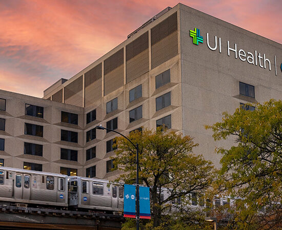
Training Affiliates Heading link
Come learn and practice in a best-in-class hospital system. Chicago campus has access to the UI Health facilities and offers exposure to a diverse patient population.

Alumni and Friends Heading link
UI COM Chicago loves to hear from alumni! Share your contact info and let us know what you’ve been up to.