Global Ophthalmology Heading link
Education Heading link
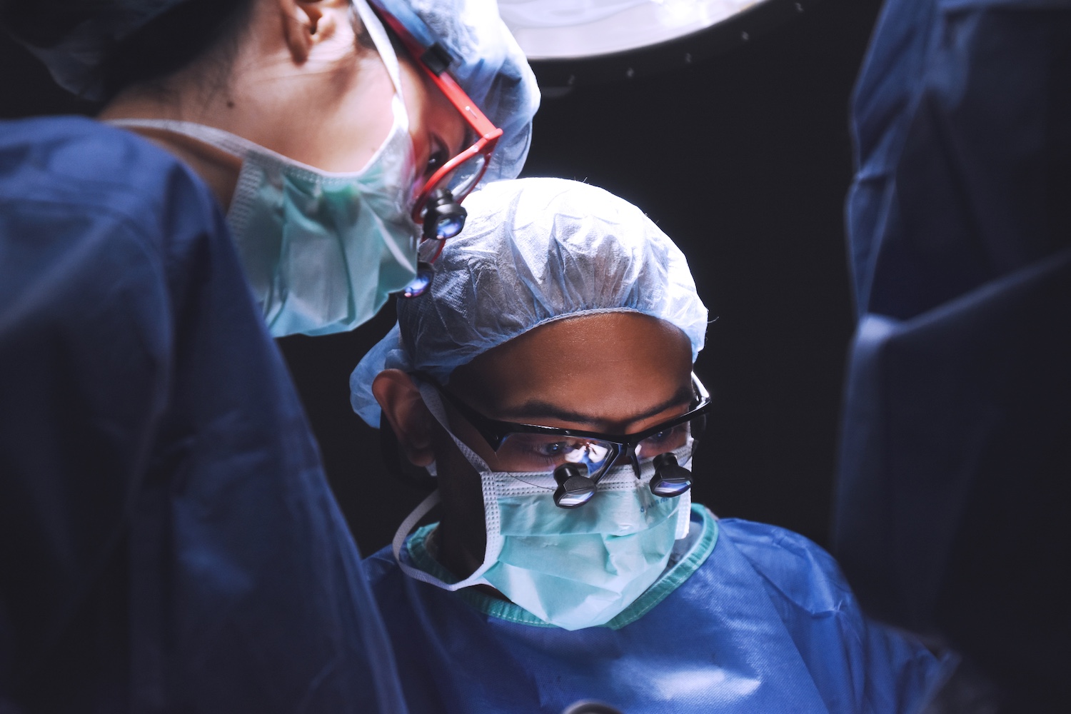
Learn more about our Residency program
The medical education ophthalmology residents receive at the Illinois Eye & Ear Infirmary will assist them throughout their career. We make sure our residents are among the best-prepared candidates for fellowship or job interviews.
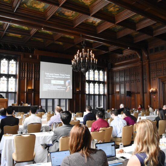
Learn about the 12 fellowship programs offered in the Department of Ophthalmology.
Each fellowship covers a different subspecialty.
Fellowship details
Medical student clerkships and workshops Heading link
The Department of Ophthalmology and Visual Sciences provides medical students with core instruction in the principles of the ophthalmologic examination.
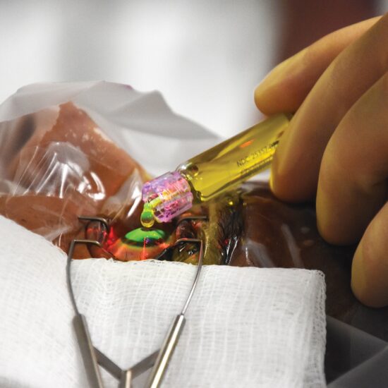
World Class Eye Care Heading link
Expert ophthalmologists at Illinois Eye & Ear at UI Health practice in a world class setting at UI Health’s new Specialty Care Building. From outpatient eye surgery to routine and complex visits, our experts provide eye care all in the same state-of-the-art building.