Clinical Psychophysics and Electrophysiology Laboratory
The research interests of this lab lie in investigations in the clinical psychophysics and electrophysiology laboratory are focused on the development of noninvasive techniques for evaluating visual function in patients with eye disease. A primary emphasis of the research in the laboratory is to establish the relationship between visual dysfunction and the underlying retinal disease process.
Overview Heading link
Ongoing research
Current investigations use psychophysical, electrophysiological, and other noninvasive procedures, in combination with information from other disciplines, such as molecular genetics. Ongoing psychophysical studies include evaluation of visual acuity, contrast sensitivity, and perimetric sensitivity. Ongoing electrophysiological studies include evaluation of rod and cone system function, as measured by the electroretinogram (ERG).
Impact
Novel, sensitive, noninvasive approaches to assessing visual function are becoming increasingly needed as gene-directed therapies and retinal cell transplantation therapies begin to enter clinical trials. In addition to assessing the efficacy of new therapeutic approaches, novel techniques for evaluating visual function can provide insight into the mechanisms underlying visual dysfunction.

Non-Invasive Methods for Assessing the Visual System Heading link
Electrophysiology
Ongoing electrophysiological studies are primarily focused on the use of the electroretinogram (ERG) to learn about retinal function in normally-sighted individuals and in patients who have acquired and inherited retinal disease.
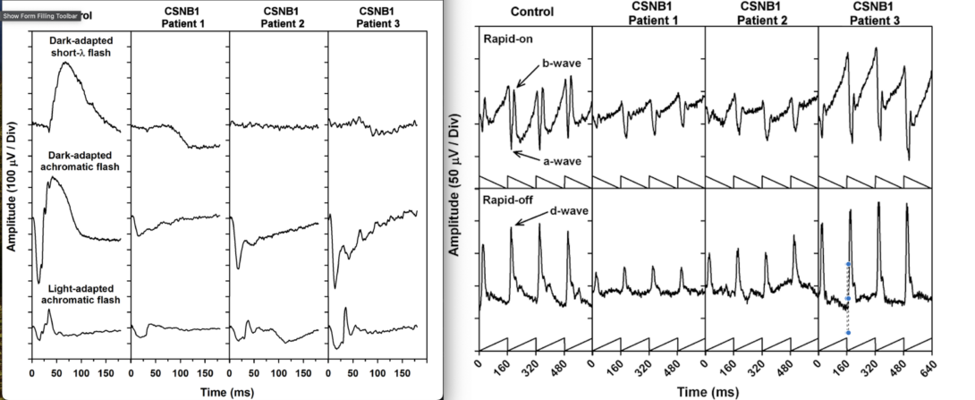
ERGs recorded in response to single flashes of light are shown in the left panel for one visually normal subject and three patients who have the complete form of congenital stationary night blindness (CSNB1). The right panel shows ERGs recorded in response to sawtooth flickering light for the same individuals. Details can be found in McAnany et al., 2013 (Oph Gen).
Recently completed and ongoing electrophysiological studies in patient populations include:
- Diabetic retinopathy
- Plaquenil toxicity
- Idiopathic intracranial hypertension
- Stargardt disease
- Congenital stationary night blindness
- X-linked retinoschisis
- Enhanced s-cone syndrome
- Achromatopsia
More Non-Invasive Methods Heading link
Psychophysics
Ongoing psychophysical studies encompass a broad range of behavioral tests including visual acuity, contrast sensitivity, and visual field perimetry. These tools permit assessing the limits of perception and performance by requiring subjects to respond to visual stimuli. The use of visual luminance noise (e.g. “TV snow”) as a tool for characterizing visual function has been of interest to us. A video of the letter ‘H’ embedded in a field of luminance noise is shown to demonstrate.
Recently completed and ongoing studies in patient populations include:
- Diabetic retinopathy
- Plaquenil toxicity
- Retinitis pigmentosa
- Choroideremia
- Leber congenital amaurosis
- Stargardt disease
Pupillometry
Ongoing pupillometric studies use the response of the pupil to a flash of light (“the pupillary light reflex;” PLR) to learn about retinal and sub-cortical neural function. We are particularly interested in measuring the PLR elicited by stimuli that can target the rod, cone, and intrinsically-photosensitive retinal ganglion cell pathways.
Watch videos of rod-meditated PLR, cone-mediated PLR and melanopsin-mediated PLR.
Recently completed and ongoing pupillometric studies in patient populations include:
- Diabetic retinopathy
- Idiopathic intracranial hypertension
- Retinitis pigmentosa
- X-linked retinoschisis
- Enhanced s-cone syndrome
- Leber congenital amaurosis
A Fourth Non-Invasive Method Heading link
Imaging
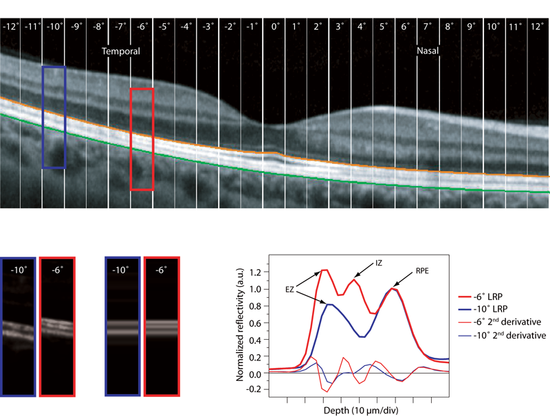
Optical coherence tomography (OCT) is a non-invasive method that permits visualizing the retina. We are interested in combining structural information, obtained by OCT, with functional measures that are under development in the lab.
Recently completed and ongoing imaging studies in patient populations include: